Microscopes are indispensable tools in various scientific and industrial fields, enabling the visualization of minute structures invisible to the naked eye. Among the diverse types available, binocular and trinocular microscopes are widely used for their enhanced depth perception and versatility. Understanding the differences between these two types is crucial for selecting the appropriate instrument for specific applications. This article aims to provide a comprehensive comparison of binocular and trinocular microscopes, covering their structure, applications, optical performance, cost, and typical user groups.

Basic Structure and Functional Differences
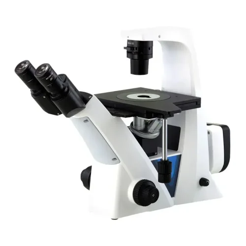
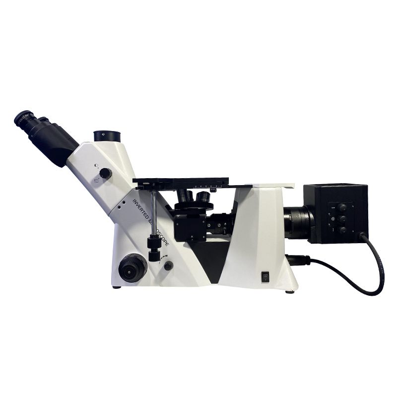
Binocular microscopes are equipped with two eyepieces to support simultaneous observation of both eyes. Their design mimics the natural perspective of the human eye and can reduce fatigue caused by long-term observation. Trinocular microscopes add a third eyepiece interface to the binocular microscope, which is specially used for external cameras, monitors or digital devices. For example, the third channel of a trinocular microscope can be directly connected to a CCD camera to achieve real-time image transmission, while a binocular microscope requires an additional adapter for photography. Below is a detailed comparison of their structural differences.
| Feature | Binocular Microscope | Trinocular Microscope |
| Number of Eyepieces | Two | Two |
| Third Port | Absent | Present, for camera/imaging device connection |
| Optical Path Design | Designed for visual observation | Includes a path for camera integration |
| External Device Compatibility | Limited to visual use | Compatible with cameras and other devices |
| Weight and Size | Generally lighter and compact | Slightly bulkier due to extra port |
| Simultaneous Viewing and Imaging | Not possible | Possible, with appropriate camera setup |
The fundamental structural difference is the presence of the third port in trinocular microscopes, enabling digital imaging capabilities. This difference dictates their respective applications and functionalities.
Application Scenarios and Professional Needs
The presence of a third port significantly expands the application scenarios for trinocular microscopes.
Binocular Microscope:
- Advantages: Simpler design, often more affordable, excellent for visual observation and routine inspections.
- Limitations: Lacks the ability to capture digital images or videos directly.
- Scalability: Limited scalability in terms of integrated imaging.
Trinocular Microscope:
- Advantages: Enables digital imaging, documentation, and sharing of microscopic observations. Ideal for research, education, and quality control where image capture is essential.
- Limitations: Typically more expensive and may require additional accessories for optimal imaging.
- Scalability: Highly scalable with various camera and software options.

Application Scenarios:
Binocular: Educational purposes, basic lab work, quality control where visual observation is sufficient.
Trinocular: Research laboratories, medical diagnostics, forensic science, material science, and industrial quality control that requires digital documentation.
Trinocular microscopes are favored in applications demanding digital imaging and documentation, while binocular microscopes are suitable for routine visual inspections and educational settings.
Optical Performance and User Experience
Both binocular and trinocular microscopes offer excellent optical performance, but the user experience can vary.
Optical Performance
- Resolution and Magnification
Binocular Microscopes:
Typically equipped with 3–4 objective lenses (e.g., 4x, 10x, 40x, 100x), offering a magnification range of 40x–1000x.
Stereoscopic vision is achievable through dual optical paths, enhancing depth perception for samples like tissues or insects. However, resolution is limited by the objective lens quality and light source (e.g., halogen or LED).
Trinocular Microscopes:
Often include 5–6 objective lenses (e.g., 2x, 10x, 20x, 40x, 60x, 100x), extending the magnification range to 20x–2000x (with oil immersion).
Advanced models support 3D imaging via specialized objectives (e.g., phase contrast, DIC), critical for analyzing transparent samples like live cells.
- Light Path Design
Binocular: Light is split equally between two eyepieces (50:50 ratio), ensuring balanced brightness for both eyes. However, attaching a camera via an external adapter (e.g., beam splitter) may reduce light intensity by 15–30%, degrading image quality.
Trinocular: Incorporates a dedicated third optical channel for imaging devices. Light distribution is adjustable (e.g., 80% to eyepieces, 20% to camera), minimizing brightness loss during simultaneous observation and recording. High-end models feature motorized light control for precision adjustments.
- Brightness and Contrast
Binocular models often rely on reflected light sources, which may struggle with low-contrast samples.
Trinocular systems frequently integrate Köhler illumination and adjustable diaphragms, enhancing contrast and uniformity. For fluorescence imaging, trinocular setups support epi-illumination with filters, critical for applications like immunofluorescence staining.

User Experience
- Ease of Operation
Binocular: Simplified design enables quick focusing (coarse/fine knobs) and stage adjustments. Ideal for rapid screening (e.g., pathology slides).
Trinocular: Requires balancing light paths between eyepieces and cameras, increasing setup complexity. Advanced models include auto-focus and motorized stages to streamline workflows. Supports real-time image sharing via HDMI/USB outputs, enabling collaborative analysis (e.g., team-based research).
- Ergonomics
Binocular eyepieces are often fixed-angle (30°–45°), forcing users to hunch over during long sessions.
Trinocular heads offer adjustable eyepieces (0°–90° tilt) and wide-field optics, reducing neck strain. Some models include ergonomic stands with height-adjustable stages.
- Workflow Integration
Binocular: Manual note-taking or sketching is required for documentation, slowing down data collection.
Trinocular: Seamless integration with imaging software (e.g., ZEN, NIS-Elements) enables automated measurements, time-lapse recording, and AI-driven analysis (e.g., cell counting).
Both microscope types provide good optical performance, but trinocular microscopes with digital imaging capabilities can enhance user experience by reducing eye strain and facilitating collaborative viewing.
Cost and Maintenance Considerations
Cost and maintenance are significant factors in microscope selection.
Purchase Cost:
Binocular microscopes are generally more affordable than trinocular microscopes due to their simpler design.
Trinocular microscopes require additional investment in cameras and imaging software, increasing the overall cost.
Maintenance Cost:
Both types require regular cleaning and maintenance of optical components.
Trinocular microscopes may require more frequent maintenance of the camera and imaging system. Replacing or repairing the light splitting prism within a trinocular microscope can be costly.
Binocular microscopes offer a lower initial cost, while trinocular microscopes come with higher upfront and potential maintenance costs due to their added complexity and imaging capabilities.

Who Prefers Binocular or Trinocular Microscopes?
The choice between a binocular and trinocular microscope is heavily influenced by the user’s specific needs and the nature of their work. Here are some typical user groups and their preferences:
Binocular Microscope Users:
- Educational Institutions (Primary and Secondary Schools, Introductory College Labs): Students and educators often utilize binocular microscopes for basic observation and learning. The simplicity and lower cost make them ideal for teaching fundamental microscopy skills.
- Hobbyists and Amateur Scientists: Individuals interested in observing insects, plants, or other small specimens for personal enrichment find binocular microscopes sufficient for their needs.
- Quality Control Technicians (Basic Inspection): In industries where routine visual inspection is required without the need for digital documentation, binocular microscopes are commonly used.
- Small Clinics and Basic Laboratories: For simple examinations that do not require digital recording, such as some parasitology or simple cell counts, binocular microscopes are adequate.

Trinocular Microscope Users:
- Research Scientists (Life Sciences, Materials Science, etc.): Researchers who need to capture high-resolution images and videos for analysis, documentation, and publication rely on trinocular microscopes.
- Medical Diagnostic Laboratories: Pathologists, cytologists, and other medical professionals use trinocular microscopes for detailed examination of tissue samples and cells, often requiring digital imaging for accurate diagnosis.
- Forensic Scientists: Forensic investigators use trinocular microscopes to analyze evidence such as fibers, hairs, and other trace materials, requiring precise image capture for documentation and analysis.
- Industrial Quality Control (Advanced Inspection): Industries requiring detailed documentation of microscopic defects or features, such as in semiconductor manufacturing or materials testing, utilize trinocular microscopes.
- Advanced Educational Settings (University Research Labs): University level researchers, and graduate students often require the digital capturing abilities of a trinocular microscope.
- Professionals in Digital Pathology and Telemedicine: Trinocular microscopes are vital for capturing and sharing digital images of tissue samples for remote diagnosis and consultation.
In summary, the primary difference between binocular and trinocular microscopes lies in the presence of a third port for camera integration. This feature significantly impacts their applications, with trinocular microscopes offering enhanced digital imaging capabilities for research and documentation, while binocular microscopes serve as cost-effective tools for routine observation and educational purposes. When selecting a microscope, you should carefully consider your specific needs, budget, and the required level of imaging capabilities. If you need some choosing suggestions, please feel free to contact the microscope supplier.


