Microscopes are the core tools for biological research, and their development history runs through the human exploration of the microscopic world of life. From Leeuwenhoek’s single microscope in the 17th century to modern super-resolution imaging systems, technological advances have continuously broken through the limits of observation. The core function of biological microscopes is to reveal the dynamic structure and function of cells, molecules, and tissues, and its application range covers basic cell biology, pathology, drug development and other fields. With advancements in technology, a wide variety of microscopes are available today, each tailored for specific applications and levels of detail. Let’s know their differences and to choose the right microscope for your needs.
What Are the Microscopes Used in Biology?
Microscopes used in biology can be broadly classified based on their source of illumination and functionality. They include light microscopes, electron microscopes, and advanced/specialized microscopes. Light microscopes utilize visible light, while electron microscopes use beams of electrons, offering significantly higher resolution. Beyond these, other specialized techniques, like atomic force microscopy and super-resolution microscopy, offer unique advantages for specific applications. Below is a detailed breakdown:
Overview Sheet of Microscope Types
| Classification | Microscope Type | Working Principle | Structure Overview | Key Features | Applications |
| Light Microscopes | Brightfield Microscope | Light passes through the sample. | Uses lenses to magnify the image. | Simple, inexpensive. | Viewing stained cells and tissues. |
| Darkfield Microscope | Oblique illumination. | Special condenser to block direct light. | Enhanced contrast, dark background. | Viewing unstained, transparent specimens, motile organisms. | |
| Phase Contrast Microscope | Exploits differences in refractive index. | Specialized optics to enhance contrast. | Visualization of live, unstained samples. | Observing live cells and their internal structures. | |
| Fluorescence Microscope | Uses fluorescent dyes or proteins. | Excitation and emission filters. | High specificity, localization of molecules. | Identifying specific molecules, visualizing cellular processes. | |
| Confocal Microscope | Laser scanning and pinhole apertures. | Laser source, pinhole apertures, detectors. | High resolution, 3D imaging. | Creating high-resolution 3D images, optical sectioning. | |
| Stereo Microscope | Provides a 3D view of larger specimens using reflected light. | Two eyepieces, dual optical paths, and a light source for surface illumination. | Low magnification (up to 50x); offers depth perception. | Dissections, surface analysis, and viewing large biological specimens. | |
| Electron Microscopes | TEM Microscope | Electrons pass through an ultrathin sample. | Electron gun, electromagnetic lenses. | Extremely high resolution. | Visualizing ultrastructure of cells and organelles. |
| SEM Microscope | Electrons scan the surface of a sample. | Electron gun, scanning coils, detectors. | 3D surface views. | Studying surface morphology and topography. | |
| Other Techniques | AFM Microscope | Probe scans the sample surface. | Cantilever, probe tip. | Nanoscale resolution. | Studying surfaces at the nanoscale, manipulating single molecules. |
| Super-Resolution Microscope | Specialized techniques to overcome diffraction limit. | Varies depending on the technique. | Higher resolution than traditional light microscopy. | Visualizing cellular structures at higher resolution. | |
| Multiphoton Microscope | Uses lasers to excite fluorophores at a specific focal point. | Laser source, specialized optics. | Imaging deep within tissues, reduced photobleaching. | Imaging thick tissues, live animal imaging. |
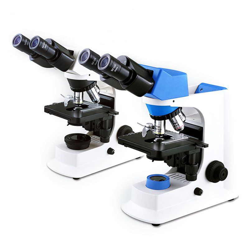
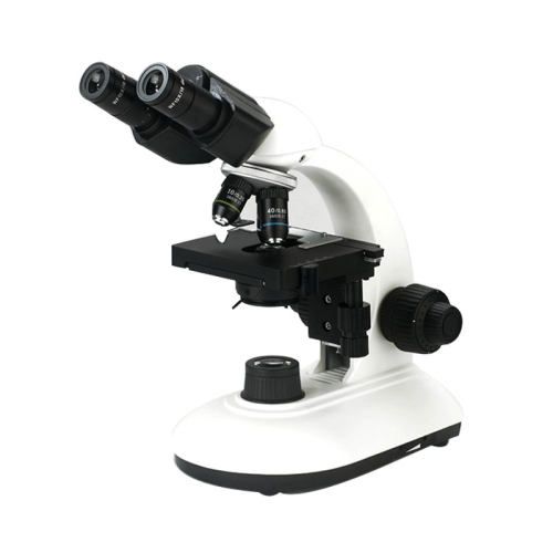
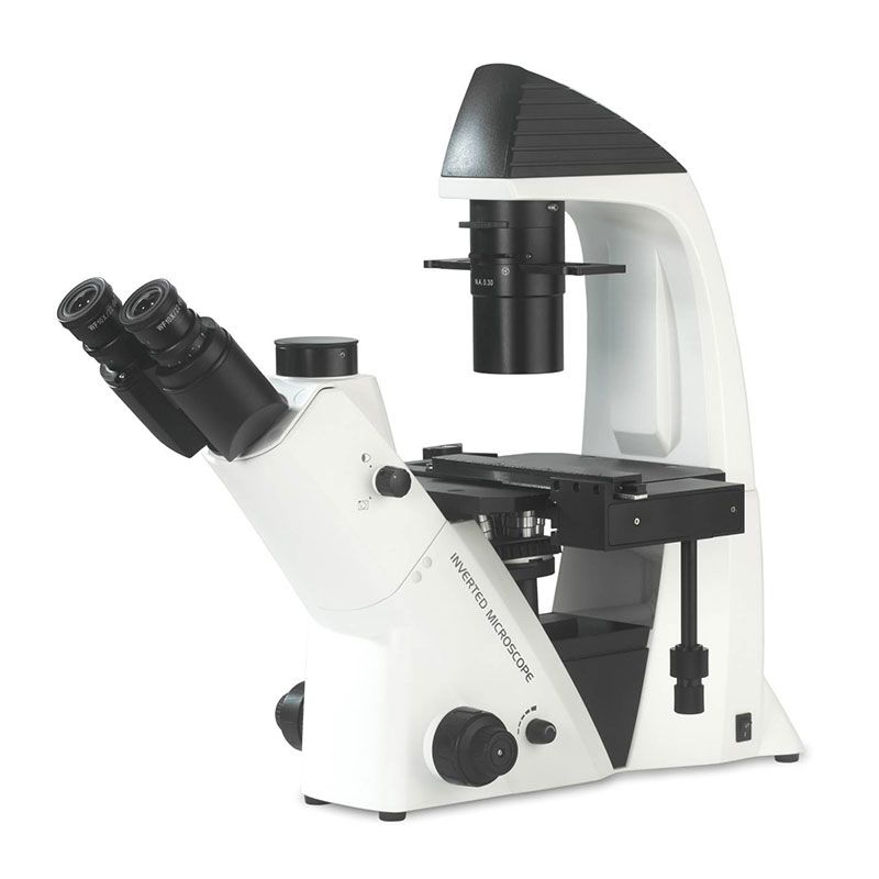
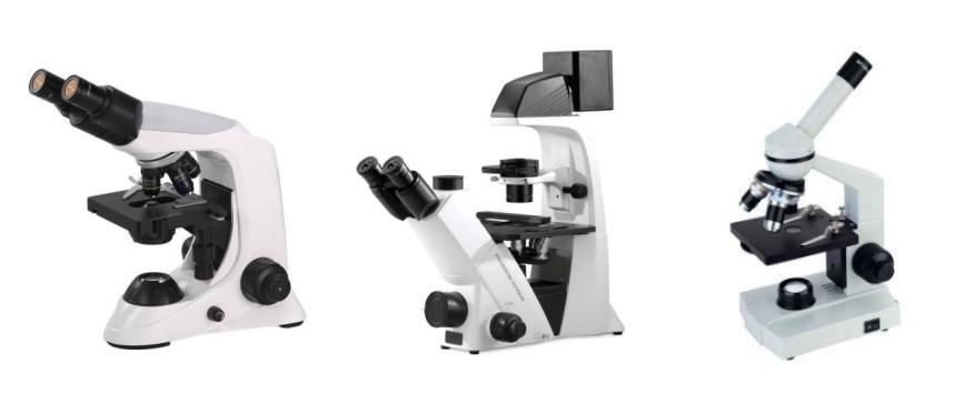
Light Microscopes
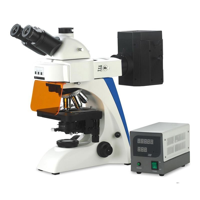
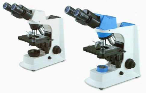
Light microscope, despite its lower resolution compared to electron microscope, remains a cornerstone of biological research due to its versatility and accessibility. Brightfield microscope, the simplest form, is ideal for viewing stained preparations. Darkfield and phase contrast microscope enhance contrast in unstained samples, making them suitable for observing live cells and microorganisms. Fluorescence microscope, with its ability to visualize specific molecules, has revolutionized cell biology. Confocal microscope allows for high-resolution 3D imaging. They are like high-powered microscopes that allow you to see very fine details within cells and tissues, even in 3D. Stereo Microscope has lower magnification and resolution compared to confocal microscopes. They are more like magnifying glasses that give you a 3D view of larger objects, making them ideal for dissections and manipulations.
Light microscope has been instrumental in countless biological discoveries, from identifying the structure of DNA to visualizing the dynamic processes of cell division. Its ease of use and affordability make it an indispensable tool in any biological laboratory.
Electron Microscopes
Electron microscope, with its use of electron beams, offers a dramatic increase in resolution, allowing for the visualization of cellular ultrastructure. TEM provides detailed images of the internal structures of cells and organelles, while SEM reveals the intricate surface topography of samples.
Electron microscope has provided invaluable insights into the fine details of cellular organization, revealing the intricacies of organelles, viruses, and macromolecules. It has been essential in fields like virology, structural biology, and materials science.
The choice of microscopy technique should take into account factors such as sample characteristics (live/fixed), resolution requirements and budget constraints.
Applications of Microscope in Biological Research
Biological microscope plays a crucial role in virtually every area of biological research:
- Cell Biology: Studying cell structure, division, signaling pathways, and organelle function.
- Microbiology: Visualizing bacteria, viruses, fungi, and other microorganisms, studying microbial interactions.
- Histology: Examining tissue structure and organization, identifying pathological changes.
- Developmental Biology: Studying embryogenesis, tissue differentiation, and developmental processes.
- Neurobiology: Mapping neural circuits, visualizing synaptic activity, studying brain structure and function.
- Genetics: Visualizing chromosomes, studying gene expression patterns.
- Pharmacology: Investigating the effects of drugs on cells and tissues.
From understanding the fundamental processes of life to diagnosing diseases and developing new therapies, microscope is an indispensable tool for biological research.
Choosing the Right Biological Microscope
Selecting the appropriate microscope depends on the specific requirements of your research or application. Consider the following factors:
- Magnification and Resolution: Determine the level of detail needed. For ultrastructures, electron microscopes are suitable, while light microscopes suffice for general observations.
- Sample Type: Identify whether you need to study live or fixed samples, stained or unstained, thin or thick specimens.
- Functionality: Opt for specialized microscopes (e.g., fluorescence or phase-contrast) for advanced imaging.
- Budget and Maintenance: High-end microscopes like TEM and AFM are expensive and require regular maintenance.
Recommended Biological Microscope Suppliers
Numerous reputable suppliers offer a wide range of biological microscopes, catering to different needs:
Olympus: Known for high-quality optical systems and innovative designs.
Leica Microsystems: Specializes in advanced imaging systems, including confocal and super-resolution microscopes.
Zeiss: Offers a diverse range of microscopes for research and clinical use.
Nikon: Renowned for ergonomic designs and precision optics.
Scopelab: Supplies various microscopes for students, teaching, and research etc. With high-performance and affordable prices.

By understanding your needs and evaluating the features of different microscopes, you can select the best tool to achieve your research goals.
Microscopes have transformed biological research, opening up new possibilities for understanding the intricate details of life. From basic light microscopes to advanced electron and confocal systems, these tools cater to a wide range of applications. By selecting the right microscope and leveraging its features, researchers can unlock new insights and contribute to scientific progress. As technology continues to evolve, the future of microscopy promises even greater capabilities and discoveries.