News
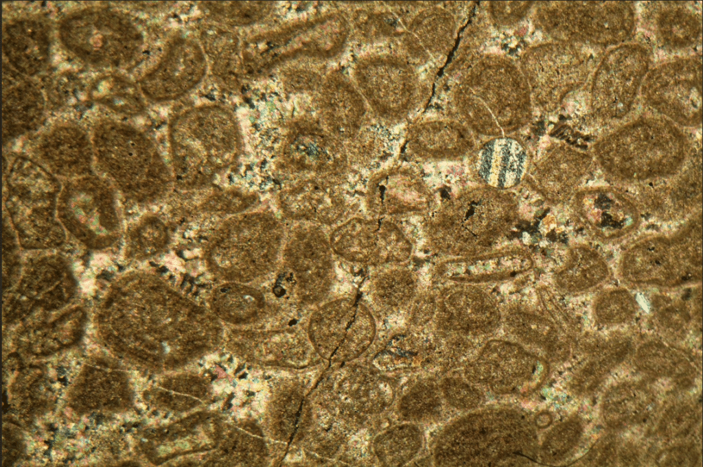
How to identify different minerals under a microscope?
When identifying minerals under a microscope, the most reliable method is not “guessing the name by color,” but using a polarizing microscope to combine color, relief, cleavage, and pleochroism under plane-polarized light with interference color, twinning, and extinction characteristics under crossed polars. Although there are thousands of minerals in nature, over 99% of Earth’s crustal…
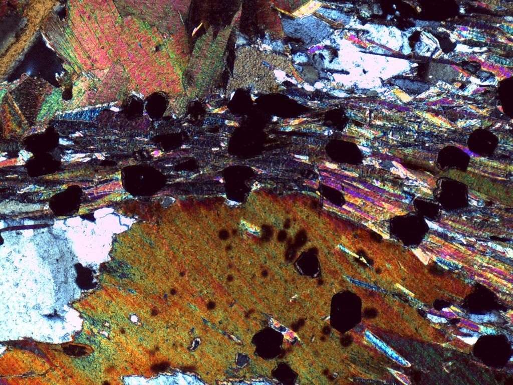
How Does an Isotropic Mineral Appear in a Microscope?
Under a polarizing microscope, an isotropic mineral appears black (extinction) under cross-polarized light (XPL) and remains black throughout a full rotation of the microscope stage. This is the main characteristic for identifying isotropic minerals. Microscope State Appearance of Isotropic Mineral Plane-Polarized Light (PPL) Transparent or colored, not necessarily black Cross-Polarized Light (XPL) Black / Extinction…
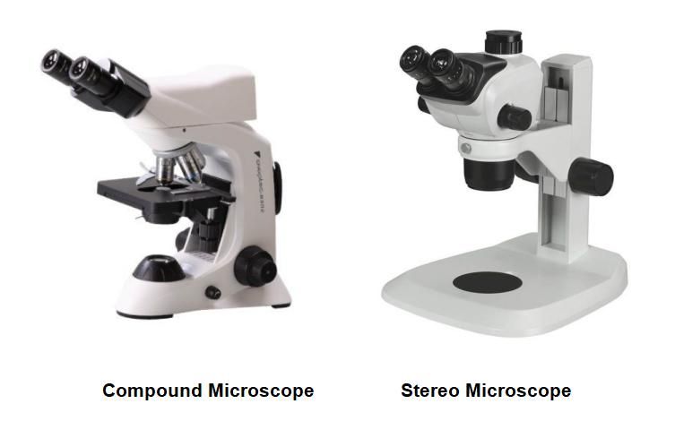
How does a stereo microscope differ from a compound microscope? (2D vs 3D)
Stereo microscopes display larger 3D objects at low magnification, allowing for observation and hands-on operation, whereas compound microscopes display very small, thin samples at high magnification, allowing for the observation of cells and fine internal structures. Features Stereo Microscope Compound Microscope Also called Dissecting microscope Biological / High-power microscope Typical magnification Low (typically 10x to…
Can You See a Plant Cell with a Light Microscope?
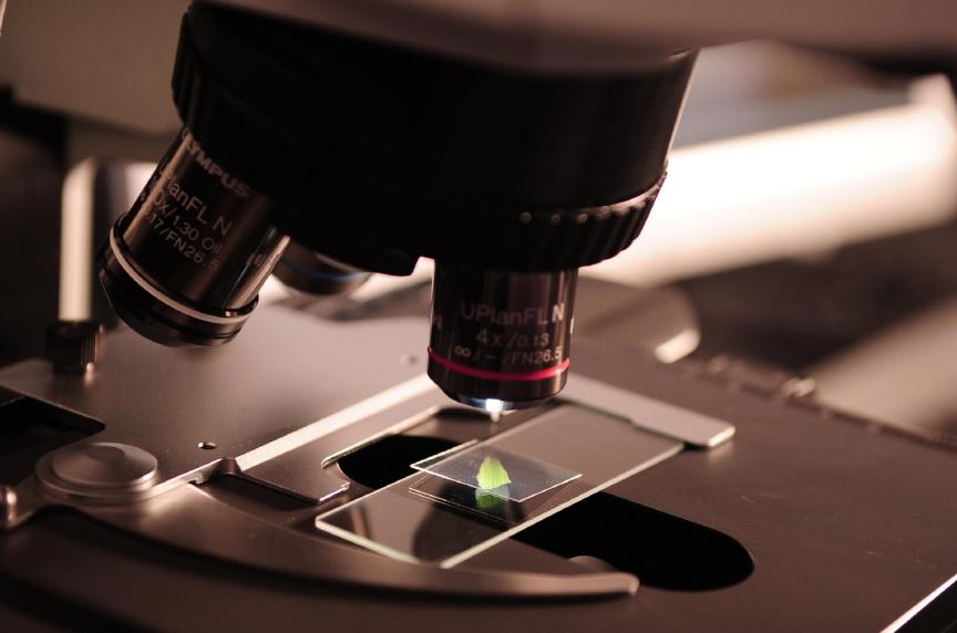
Yes, you can easily observe plant cells using a standard light microscope. It is especially suitable for thin specimen samples such as onion skin, moss leaves, or Elodea (waterweed). Typically, you can see the cell wall, cell membrane, cytoplasm, nucleus, large central vacuole, and chloroplasts. What Can You See Under a Light Microscope? Under a…

Why Are Stains Used When Observing Cells Under the Microscope?
Stains are used to make cells and their internal structures easier to observe. Under a standard light microscope, most cells and their internal components are naturally colorless and translucent (almost entirely transparent). Unmodified living cells are a bit like trying to spot a clear jellyfish in clear water: light passes right through them with almost…

Can You See a Plant Cell with a Light Microscope?
Yes, plant cells can be easily observed using a light microscope. A standard compound light microscope (40x to 400x) can clearly show the cell wall, chloroplasts (in green tissues), and large central vacuoles (such as a thin layer of onion epidermis or green leaves of aquatic plants). However, only certain structures are visible; finer organelles like…

Which Two Microscopes Generate Three-Dimensional Images for Semiconductor Inspection?
In semiconductor manufacturing, component features have shrunk into micro- and nanometer scales, with architectures transitioning from 2D planes to 3D structures (such as multi-layer stacks, FinFETs, and GAA transistors). Traditional 2D planar imaging cannot measure critical vertical metrics like solder bump height or trench depth. Extracting height, depth, and volume accurately requires 3D microscopy. In…

What Microscope Is Best for PCB Inspection in SMT & EMS Manufacturing?
In Surface Mount Technology (SMT) and Electronics Manufacturing Services (EMS) facilities, the optimal equipment strategy is: deploy 60 FPS @ 4K/1080P high-frame-rate digital microscope systems with cross-polarization at SMT inline inspection and First Article Inspection (FAI) stations to eliminate motion lag and reflections, boosting UPH and reducing fatigue; deploy trinocular stereo boom microscopes at dedicated…

Where can I find a 200mm / 230mm platen metallographic polisher?
If you are looking for a metallographic grinder-polisher with a 200 mm or 230 mm working wheel (platen/sandpaper diameter), the primary procurement channels include: First, what are the main classifications of metallographic polishers and their differences in sample applications? Type Key Features & Configuration Sample Applications & Best Use Cases Manual Grinder-Polisher Single or dual…

How to Automate the Metallographic Polishing Process?
To automate the metallographic polishing process, the most direct and effective solution is to adopt a commercial Fully Automatic Metallographic Polisher. Achieving true automation relies on locking down the four critical parameters that govern sample quality—force, speed, time, and lubricant/slurry dosing. Using a Fully Automatic Metallographic Polisher: The Most Efficient & Direct Approach To automate…
Can Brinell Hardness Testing Be Used on Corroded Metal?
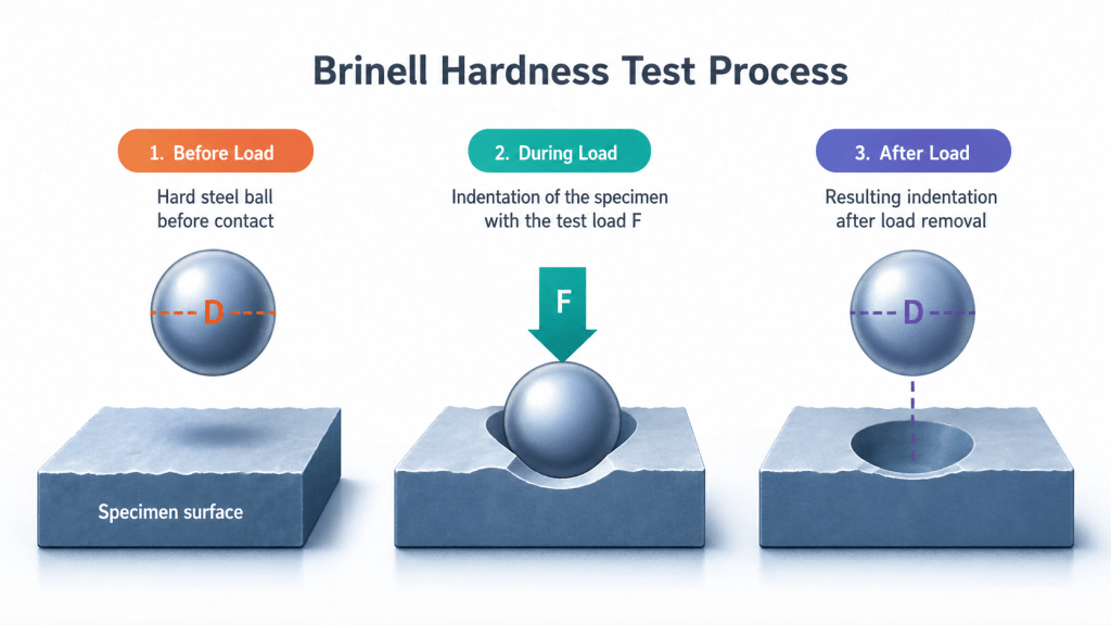
Well, yes. Although Brinell hardness testing can be used on corroded objects, it absolutely cannot be performed directly on the raw, original corroded surface. You must use surface preparation like grinding to clear away the top corrosion products and expose the undamaged base metal inside. Testing directly on rust, mill scale, pitting, or a loose…
What is Another Way to Test Hardness of Metals?
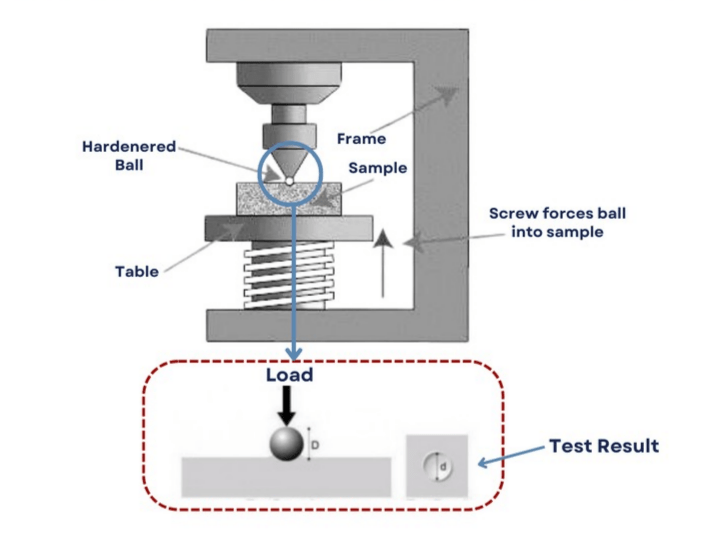
When conventional hardness testers (such as Rockwell, Brinell, or benchtop Vickers units)—and even portable electronic devices like Leeb or Ultrasonic Contact Impedance (UCI) tools—are unavailable or impractical, alternative field-tested methods can be used to evaluate metal hardness on the shop floor. Most Popular: File Testing (Scratch Method) — Best for Heat Treatment (Quenching/Tempering) On-Site Inspections…