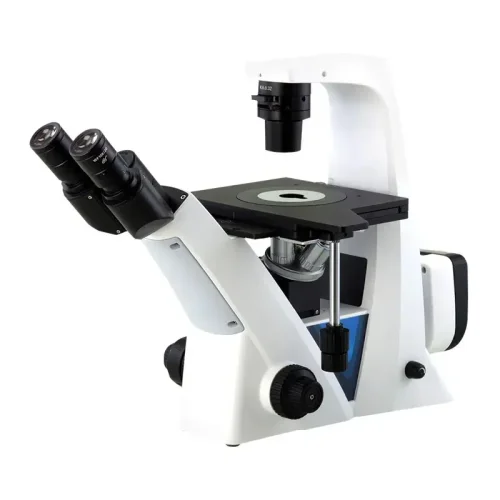
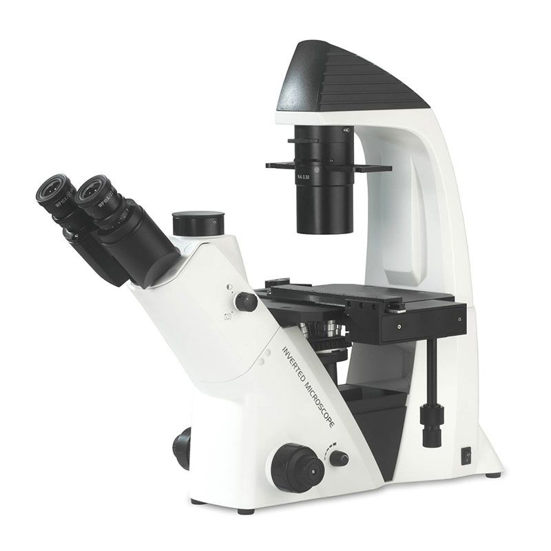
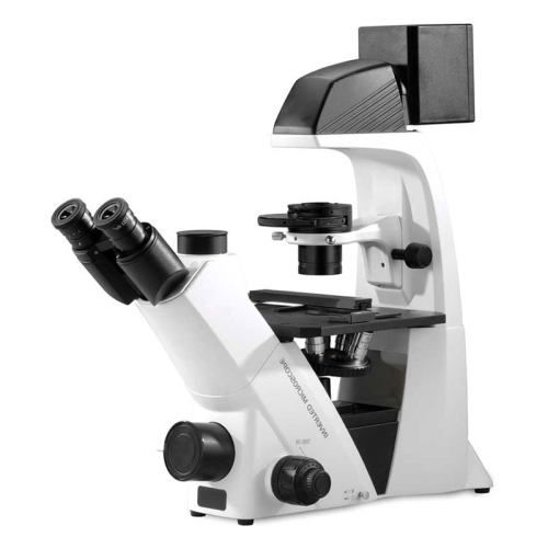
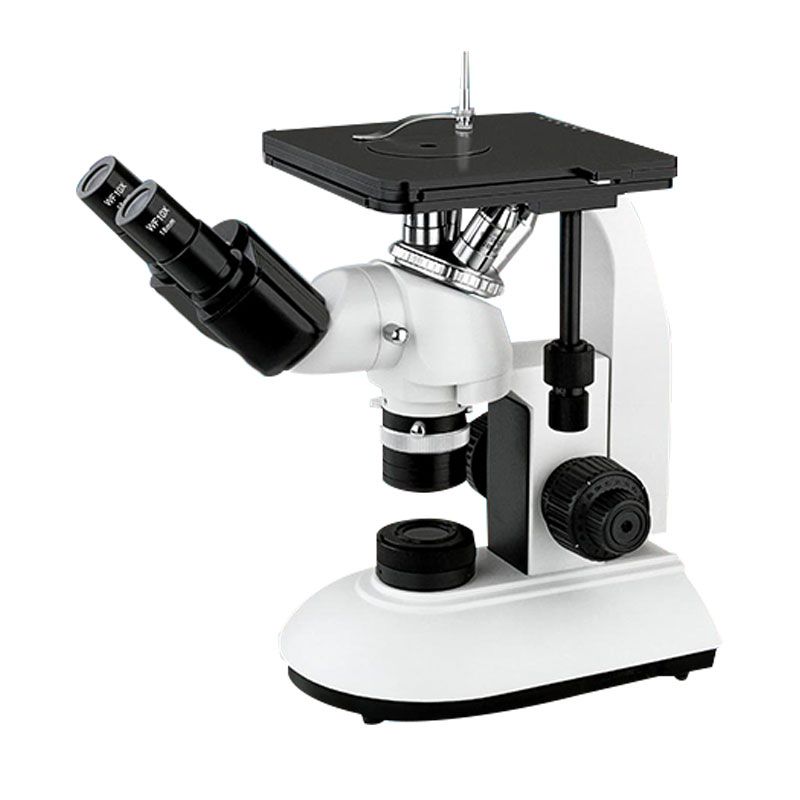
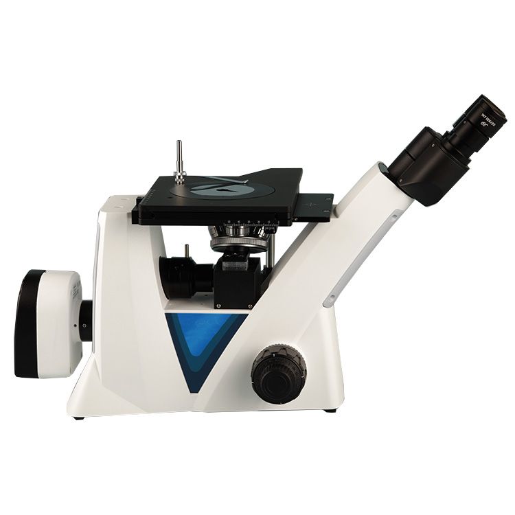
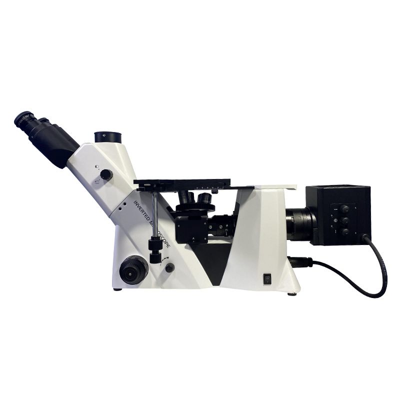
Inverted biological microscopes have become indispensable tools in modern scientific research. Unlike traditional upright microscopes, these instruments are designed with the objective lens below the specimen, making them particularly suitable for observing living cells and organisms in culture dishes. This article explores the top features of advanced inverted biological microscopes, shedding light on how these innovations are enhancing research capabilities and driving scientific discoveries.
1. Enhanced Optical Systems
High-Resolution Imaging
One of the most significant advancements in inverted biological microscopes is the development of high-resolution optical systems. Modern microscopes employ advanced lens technologies that significantly improve image clarity and resolution. This allows researchers to observe minute cellular structures and processes with exceptional detail. For example, the use of apochromatic lenses minimizes chromatic aberrations, ensuring that images are sharp and color-accurate.
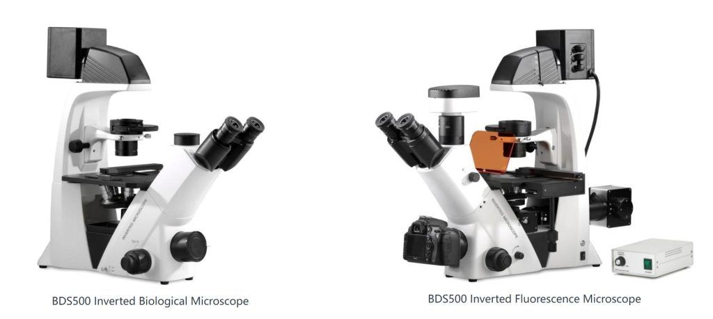
Fluorescence Capabilities
Fluorescence microscopy has become a cornerstone technique in biological research. Advanced inverted microscopes are equipped with sophisticated fluorescence capabilities, enabling the visualization of specific cellular components using fluorescent dyes and proteins. These microscopes often include multiple fluorescence channels, allowing for multi-color imaging and the study of complex cellular interactions.
2. Automated and Motorized Functions
Precision Control
Automation and motorization have revolutionized microscopy, bringing precision control to the forefront. Advanced inverted microscopes feature motorized stages and focus mechanisms, allowing for precise positioning and focusing of samples. This is particularly beneficial for experiments requiring high repeatability and accuracy, such as time-lapse studies.
Automated Imaging
The integration of automated imaging systems has streamlined the data acquisition process. These microscopes can be programmed to perform automated scanning and imaging, reducing the need for manual intervention. This capability is essential for high-throughput screening applications, where large volumes of samples need to be analyzed efficiently.
3. Advanced Imaging Techniques
Confocal and Multi-Photon Microscopy
Advanced inverted microscopes are often compatible with confocal and multi-photon microscopy techniques. Confocal microscopy provides high-resolution, three-dimensional images by focusing on a specific plane within the specimen, reducing background noise. Multi-photon microscopy allows for deep tissue imaging, making it ideal for studying thick specimens and living tissues.
Live Cell Imaging
Live cell imaging has become crucial for understanding dynamic biological processes. Advanced inverted microscopes are equipped with features that support live cell imaging, such as environmental control chambers that maintain optimal conditions for cell viability. This enables researchers to conduct long-term studies of cellular behaviors and interactions in real-time.
4. Improved Ergonomics and User Interface
User-Friendly Interface
Ease of use is a critical factor in scientific instrumentation. Modern inverted microscopes come with user-friendly interfaces, including touchscreen controls and intuitive software. These interfaces simplify the operation of complex microscopy functions, making it easier for researchers to focus on their experiments rather than the technical aspects of the equipment.
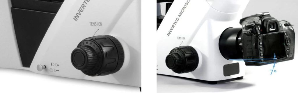
Ergonomic Design
Ergonomics is another area where significant improvements have been made. Advanced inverted microscopes feature adjustable components that can be tailored to the user’s comfort. This is particularly important for reducing fatigue during extended periods of microscope use, thereby enhancing productivity and reducing the risk of repetitive strain injuries.
5. Integration with Digital and Computational Tools
Digital Imaging and Analysis
Digital imaging technology has transformed microscopy. High-resolution digital cameras integrated into advanced inverted microscopes capture detailed images that can be easily stored, shared, and analyzed. Sophisticated software tools are available for image analysis and data management, enabling researchers to quantify and interpret their observations with greater accuracy.
AI and Machine Learning Integration
The integration of artificial intelligence (AI) and machine learning (ML) is a cutting-edge development in microscopy. AI algorithms can assist in image recognition and analysis, identifying patterns and anomalies that might be missed by the human eye. This technology holds immense potential for accelerating research and improving the accuracy of data interpretation.
6. Versatility and Adaptability
Modular Design
The modular design of advanced inverted microscopes offers unparalleled versatility. Researchers can customize and upgrade their microscopes with various components, such as different objective lenses, illumination systems, and imaging modules. This adaptability ensures that the microscope can evolve with the changing needs of the research.
Compatibility with Multiple Techniques
Advanced inverted microscopes are designed to be compatible with a wide range of microscopy techniques. This includes brightfield, phase contrast, differential interference contrast (DIC), and more. The ability to switch between techniques makes these microscopes valuable tools for multi-disciplinary research, allowing scientists to apply the most appropriate method for their specific study.
Conclusion
Advanced inverted biological microscopes are at the forefront of scientific innovation, offering a host of features that enhance research capabilities. From high-resolution optical systems and fluorescence capabilities to automated functions and AI integration, these microscopes are transforming how researchers observe and understand biological processes. As technology continues to evolve, we can expect further advancements that will push the boundaries of what is possible in microscopy, driving new discoveries and insights in the life sciences.